Pulmonary Embolism: Causes, Symptoms, and Management Strategies
This article explains pulmonary embolism (PE), including its causes, symptoms, risk factors, diagnostic methods, and treatment options. Prompt medical intervention is crucial to prevent severe complications. Prevention strategies focus on managing risk factors like blood clots, obesity, and prolonged bed rest. Early diagnosis and appropriate treatment significantly improve outcomes, making awareness and prevention essential for at-risk individuals.

Pulmonary Embolism: Causes, Symptoms, and Management Strategies
Pulmonary embolism (PE) happens when a blood clot blocks one of the arteries in the lungs, often stemming from deep vein thrombosis in the legs. This blockage hampers blood flow and can be life-threatening if not addressed swiftly. In rare cases, clots may originate elsewhere. Prompt medical care is crucial for reducing risks. Preventative steps include controlling leg blood clots and early detection to prevent serious complications.
Common signs of PE
Symptoms vary depending on clot size and lung involvement. Typical symptoms include sudden difficulty breathing, chest pain similar to a heart attack, coughing with blood streaks, dizziness, rapid heartbeat, sweating, fever, skin discoloration, leg pain, and swelling. Individuals with existing heart or lung issues may experience more severe symptoms.
Causes of pulmonary embolism
PE typically results from a blood clot blocking a lung artery, most often originating from deep vein thrombosis in the legs. Clots may also form from air bubbles, fat, or tumor parts, leading to serious issues like pulmonary infarction, where oxygen supply to tissues is severely reduced.
Factors increasing PE risk
While anyone can develop blood clots, certain factors heighten the risk. These include family history, heart ailments, cancers (particularly lung, ovarian, or pancreatic), recent surgeries, extended bed rest, smoking, obesity, pregnancy, and hormone therapies. Awareness and preventative measures are vital for high-risk individuals.
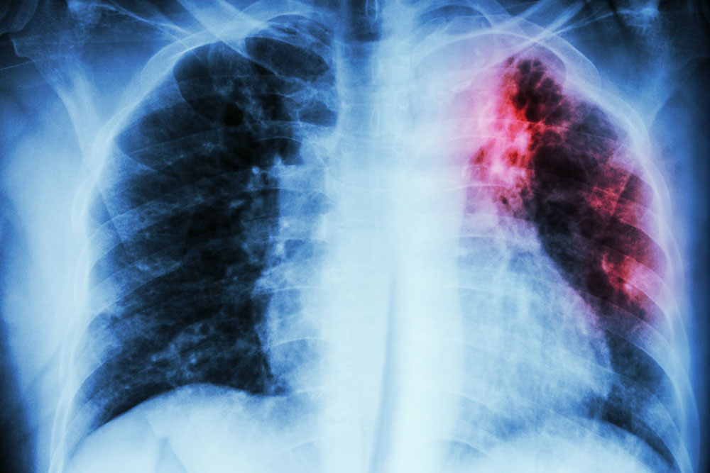
PE diagnosis methods
Diagnosis involves reviewing symptoms, physical exams, and imaging tests such as chest X-ray, ECG, MRI, CT scan, pulmonary angiography, ultrasound, or venography. Blood tests like D-dimer assist in detecting clots, helping confirm the diagnosis.
Treatment options
The treatment approach depends on the clot's size and location. Early detection often allows for anticoagulant drugs or thrombolytics. Severe cases may require surgical procedures such as clot removal, vena cava filter placement, or emergency bypass surgery. After treatment, managing underlying causes with blood thinners, compression stockings, and leg exercises helps prevent recurrence.