Comprehensive Guide to Bone Density Testing and Its Significance
This article provides an in-depth overview of bone density assessments, highlighting their importance in diagnosing osteoporosis and assessing fracture risk. It explains who should get tested, the procedure involved, and what the results indicate, emphasizing the critical role of early detection in maintaining bone health across different populations.
Comprehensive Guide to Bone Density Testing and Its Significance
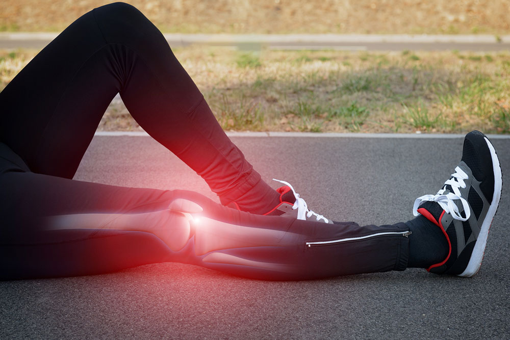
Osteoporosis is a disease where bones become fragile and more prone to fractures, particularly affecting the spine, wrist, and hips. Although more common in women, men are at risk too. In the United States, around 10 million people (8 million women and 2 million men) suffer from osteoporosis, with an additional 34 million at risk due to low bone mass, called osteopenia.
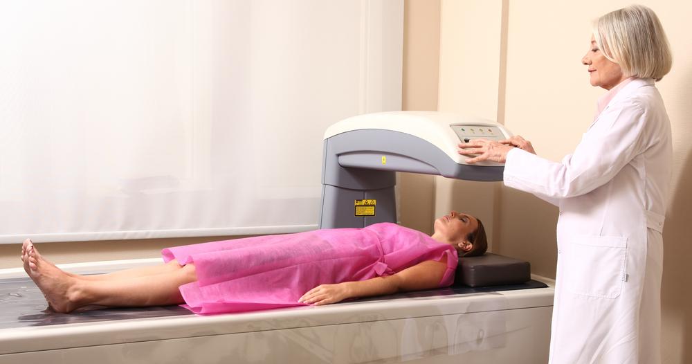
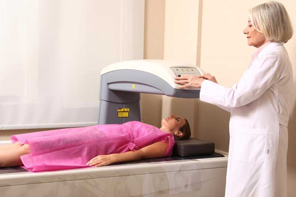
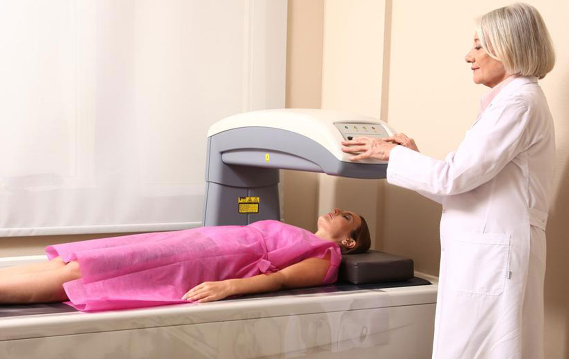
A bone density scan is a quick, painless test used to identify osteoporosis and evaluate fracture risk. It also helps monitor treatment progress. Medical professionals may recommend these scans based on individual risk factors.
Height loss: Losing more than 1.6 inches may signal spinal fractures linked to osteoporosis.
Bone fragility: Weaker bones are more susceptible to breaks.
Medication effects: Long-term use of certain drugs can impair bone formation, increasing osteoporosis risk.
Organ transplants: Transplant operations can impact bone health and elevate osteoporosis chances.
Hormonal shifts: Postmenopausal women experience reduced estrogen levels, while treatments for cancer can lower testosterone, both contributing to osteoporosis risk in men and women.
Low-dose X-ray scans identify decreases in bone mineral density, focusing mainly on the hips and spine. If hip scans are not feasible, forearm measurements are used, especially in hyperthyroidism cases. For individuals under 60, hip scans are generally recommended.
These tests measure bone density in areas like the total body, spine, and hips, while peripheral devices assess density in the heel, shin, wrist, and fingers. The procedure takes about 15 minutes, with the patient lying on a table for 5-8 minutes. Detailed 3D images assist in evaluating age-related bone health and other conditions beyond osteoporosis.