Comprehensive Guide to Diagnosing Multiple Sclerosis
This comprehensive article explains the diagnostic process for multiple sclerosis, highlighting key criteria like dissemination in space and time, advanced imaging techniques, and vital tests such as MRI, lumbar puncture, and OCT. It emphasizes early detection's importance and guides patients on understanding the evaluation procedures involved in confirming MS diagnosis for effective management.

Comprehensive Guide to Diagnosing Multiple Sclerosis
Multiple sclerosis (MS) is a neurological autoimmune condition where the immune system attacks the myelin sheath surrounding nerve fibers. This disrupts nerve communication between the brain and body. Early diagnosis enables better management, requiring various tests to rule out other similar conditions. Accurate detection relies on specific diagnostic criteria and advanced imaging techniques to track disease progression.
Diagnostic Approach
The McDonald criteria serve as the main diagnostic framework, utilizing MRI scans, cerebrospinal fluid analysis, and clinical evaluation to identify demyelination patterns characteristic of MS.
Dissemination in Space (DIS)
This assesses lesion distribution across multiple central nervous system areas, confirmed via MRI scans, reflecting the widespread nature of MS.
Dissemination in Time (DIT)
Evidence of lesions appearing at different times, confirmed through follow-up MRI, shows disease progression. Repeated neurological episodes spaced by at least 30 days also support DIT.
The criteria have been refined with advanced tools for earlier and precise MS diagnosis.
Diagnostic Methods
Since no single test definitively confirms MS, a combination of evaluations is essential for accurate diagnosis and understanding disease severity to guide treatment strategies.
1. Neurological and Medical Assessment
Detailed medical history, neurological examinations measuring reflexes, coordination, strength, vision, and sensory function help identify abnormal signs suggestive of MS.
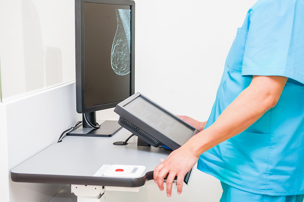
2. MRI Scans
MRI imaging highlights brain and spinal cord lesions associated with MS, with bright spots indicating demyelination. Follow-up imaging monitors disease course and therapy response.
3. Lumbar Puncture (Spinal Tap)
This procedure analyzes cerebrospinal fluid for immune markers like oligoclonal bands and elevated IgG, supporting MS diagnosis when MRI is inconclusive.
4. Blood Testing
Blood tests exclude other conditions such as infections or autoimmune diseases that have similar symptoms to MS.
5. Optical Coherence Tomography (OCT)
OCT provides detailed retinal images; thinning of the retinal nerve fiber layer indicates optic neuritis, an MS-associated condition, aiding early detection.
Important Note: The information provided is for educational purposes only and should not replace professional medical advice. Consult healthcare professionals for proper diagnosis and treatment.