Ultimate Guide to Early Detection Tests for Cancer
This comprehensive guide details essential early cancer detection tests, including imaging techniques like MRI and CT, endoscopy, biopsies, and cytology. Early diagnosis is key to effective treatment, and understanding these methods helps both healthcare providers and patients stay proactive. The article emphasizes the importance of screening programs to improve outcomes and reduce healthcare costs by catching cancer at manageable stages. Learn about the most reliable diagnostic approaches for different types of cancer and how they contribute to timely care.
Various Approaches for Prompt Cancer Diagnosis
Early identification of cancer significantly boosts treatment success and patient survival. Routine screening facilitates detection at initial stages, reducing discomfort and healthcare costs. The American Cancer Society offers key recommendations on screening methods that both healthcare professionals and individuals should know. Here are some principal tests used to detect cancer early on.
Imaging Technologies
Medical imaging enables visualization of internal organs and tissues. Techniques like ultrasound, X-rays, and advanced imaging modalities assist in locating tumors, measuring their dimensions, and understanding spread, aiding in diagnosis and treatment planning.
Computed Tomography (CT) Scans
CT scans are painless, non-invasive procedures capturing detailed cross-sectional images of bones and soft tissues. They help determine tumor size and position and are valuable for guiding therapies like heat-based tumor destruction.
Magnetic Resonance Imaging (MRI)
MRI uses magnetic fields and radio waves to produce high-resolution images of internal organs and soft tissues. Contrast dyes like gadolinium enhance visibility, especially useful for detecting metastases and identifying tumor origins. Patients are scanned while lying in a cylindrical magnet for precise diagnosis.
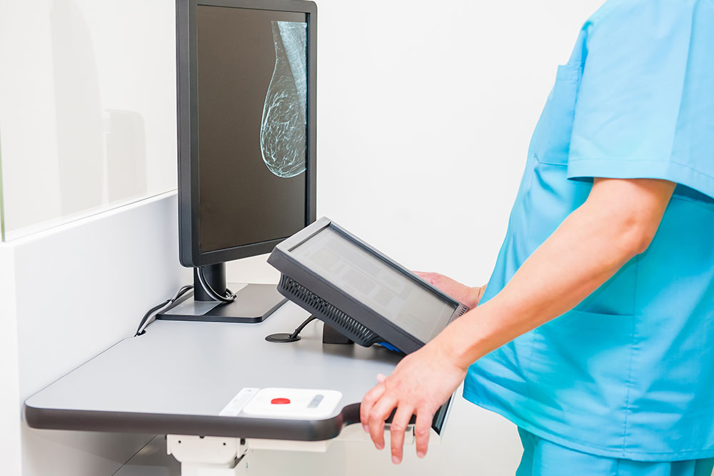
X-ray and Contrast Imaging
X-ray imaging and contrast studies help visualize internal structures and identify abnormalities. Mammography, a specialized breast X-ray, is essential for breast cancer screening, while contrast agents like barium improve imaging of the gastrointestinal system.
Nuclear Medicine Scans
This approach involves radioactive tracers that emit low-dose radiation to produce functional images showing tissue activity. Tumor sites absorb differing amounts of tracers, aiding in localization. Often used in conjunction with other tests for comprehensive analysis.
Ultrasound Diagnostics
Ultrasound employs sound waves to generate real-time images of organs and blood flow. It's particularly helpful for detecting tumors hidden on X-rays or distinguishing cysts from solid masses. Safe and quick, it has limitations in imaging air-filled or bony areas.
Endoscopic Examination
Endoscopy involves inserting a flexible tube with a camera into the body to directly view internal organs. This method aids in diagnosing cancers such as colorectal and esophageal tumors. During procedures, treatments like laser ablation can also be performed.
Tissue Sampling (Biopsy)
A biopsy confirms cancer diagnosis by extracting tissue samples using techniques like fine-needle aspiration, often guided by imaging. The collected tissue is analyzed microscopically to detect malignancy and evaluate tumor features.
Cell Tests (Cytology)
Cytology entails examining individual cells from biopsies, scrapings, or fluids like urine or sputum. It assists in diagnosing cancers, infections, and other conditions, including Pap smears for cervical cancer screening.