Advancements in Orthopedic Surgery: Key Developments and Innovations
This article explores key advancements in orthopedic surgery, highlighting minimally invasive techniques, innovative imaging technologies, and their impact on patient recovery. It traces the evolution from early arthroscopic procedures to modern diagnostic tools, emphasizing improvements that benefit both patients and surgeons.

Advancements in Orthopedic Surgery: Key Developments and Innovations
Recent progress in orthopedic treatment emphasizes minimally invasive techniques that target fewer tissues, reducing infection risks and tissue trauma, and promoting faster healing. These innovations significantly improve patient recovery experiences.
Historical Progress in the 20th Century
At the turn of the 20th century, technological limitations made it difficult to produce small surgical instruments. By the 1950s, pioneers like Dr. Masaki Watanabe pioneered arthroscopic procedures with the invention of the first arthroscope. Dr. Robert W. Jackson brought these methods to the US in the 1960s, initially facing skepticism. Early uses focused on sports injuries such as meniscus tears and ligament repairs, with challenges related to limited space and tool maneuverability.
Patients benefited from reduced bleeding, less pain, and quicker returns to normal activity, leading to broader adoption of arthroscopy across multiple joints. The addition of television monitors further enhanced the technique, enabling visualization of joints like the temporomandibular joint for more complex procedures.
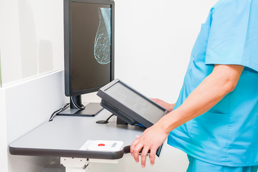
Simultaneously, breakthroughs in imaging technology—including MRI and 3D imaging—transformed diagnostics. Better ultrasonography provided clearer visualization of soft tissues, facilitating precise surgical planning and improved results.
Disclaimer:
This article covers various topics with insights aimed at informing readers. While efforts are made to ensure accuracy, the content may not cover all aspects and does not assume responsibility for external sources or potential discrepancies. Some details or offers may vary.