Essential Steps for Rheumatoid Arthritis Diagnosis
Learn about the key steps involved in diagnosing rheumatoid arthritis, including symptoms, blood tests, and imaging techniques essential for accurate detection and monitoring of the disease.
How Rheumatoid Arthritis Is Diagnosed
Rheumatoid arthritis (RA) is an autoimmune disorder resulting in joint inflammation. Here's an overview of the diagnosis process.
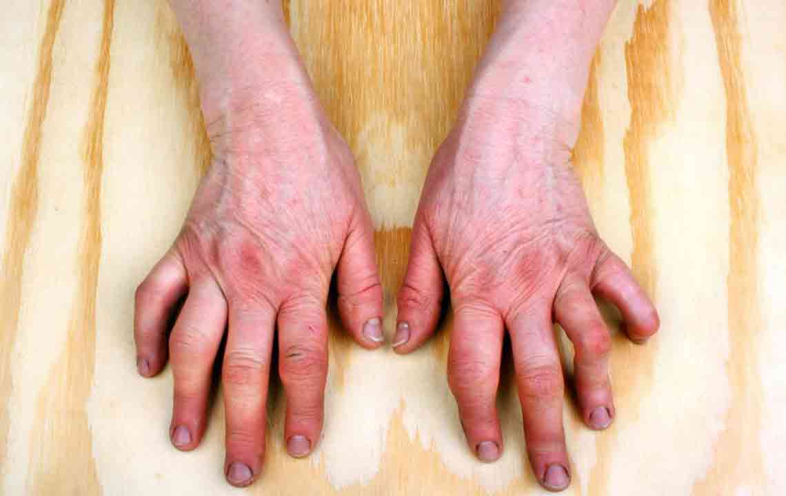
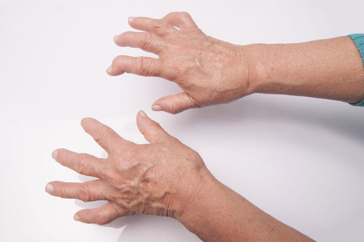
RA mostly targets small joints like those in the hands and feet, affecting approximately 1.5 million people in the U.S. If untreated, it can cause cartilage damage and deformities. It is more common in women and usually appears after 40 years old. Besides joints, RA can also involve organs such as eyes, blood vessels, skin, and lungs.
Symptoms include fatigue, fever, morning stiffness, swollen joints, pain, and weight changes. Symptoms tend to impact the same joints on both sides of the body, with severity differing among individuals. Symptoms can flare up and then subside. If untreated, RA can lead to joint deformity over time.
Since the cause of RA remains unknown, therapies focus on alleviating pain and inflammation. Accurate diagnosis is crucial before starting treatment.
RA diagnosis can be complex since other conditions show similar symptoms. No single definitive test exists. Typically, physical exams check for swelling, redness, warmth, muscle strength, and reflexes. Further blood work and imaging studies confirm the diagnosis, including:
Blood Tests
Blood tests offer clues but do not definitively diagnose RA. Key tests include:
C-Reactive Protein (CRP)
CRP measures inflammation in the liver's produced proteins. Elevated levels indicate inflammation but don't confirm RA.
Rheumatoid Factor (RF)
RF detects antibodies common in RA patients. A positive RF supports diagnosis but isn't exclusive to RA, and some patients test negative.
Complete Blood Count (CBC)
CBC evaluates blood components, revealing anemia which is prevalent in RA, but anemia alone doesn't confirm RA.
Imaging Studies
Imaging reveals joint damage and inflammation to assist diagnosis and monitor disease progression. Common methods include:
Ultrasound – Utilizes sound waves to produce joint images.
X-Ray – Uses radiation to visualize joint structures.
MRI – Employs magnetic fields and radio waves for detailed imaging.
These tools are essential for confirming RA and tracking its development.
Note:
Our platform provides diverse, practical information across various topics. While we strive for accuracy, this content is not a substitute for professional medical advice. Always consult healthcare providers for diagnosis and treatment tailored to your condition.